Enamel is the highly mineralized outer layer of the tooth, providing protection against the physical and chemical challenges of the oral environment. Despite its hardness, enamel can undergo mineral loss when exposed to acidic conditions. When these early changes remain non-cavitated, the oral environment can also support mineral recovery through remineralization.
This balance between demineralization and remineralization is an important area of preventive dentistry research. It has also led to interest in dentifrices containing bioactive materials that can interact with enamel and support mineral recovery.
One such material is calcium sodium phosphosilicate (CSPS), a bioactive glass that has been investigated in dentistry for applications including dentin hypersensitivity and enamel remineralization.
A 2017 study by Matsuyoshi and colleagues examined the effect of a dentifrice containing 5% calcium sodium phosphosilicate on enamel remineralization using optical coherence tomography (OCT). The study provides useful experimental evidence for understanding how CSPS-containing dentifrice may affect demineralized enamel. However, because the study was conducted on bovine enamel specimens under laboratory conditions, its findings should be interpreted as in-vitro evidence rather than direct evidence of clinical outcomes in patients.
Enamel Demineralization and Remineralization Dynamics
Enamel is composed predominantly of mineral and is continuously exposed to changes in the oral environment.
Acidic conditions can cause minerals to leave the enamel structure, resulting in demineralization. Saliva, on the other hand, contains ions that can contribute to the natural remineralization process.
The condition of enamel therefore reflects a dynamic balance between mineral loss and mineral gain.
When mineral loss occurs at an early stage and the enamel surface remains intact, supporting remineralization is an important principle in preventive dental care.
How Mineral Loss Changes Enamel Structure
During demineralization, mineral is lost from the enamel structure. This can produce changes within the enamel that may be detected through changes in its optical and physical properties.
Early enamel lesions may not present as visible cavities. Instead, mineral loss can occur beneath or within the enamel surface while the external surface remains relatively intact.
Understanding these structural changes is important because early intervention focuses on controlling the factors responsible for mineral loss and supporting the natural repair potential of the enamel.
Why Remineralization Is Clinically Relevant
Remineralization is important because early mineral loss is not necessarily an irreversible process.
The availability of calcium, phosphate and other relevant ions in the oral environment can contribute to mineral deposition. Fluoride also plays an established role in enamel remineralization and caries prevention.
This has encouraged research into remineralizing dentifrices containing additional bioactive ingredients, including calcium sodium phosphosilicate.
The clinical relevance of these materials lies primarily in their potential contribution to preventive approaches before an early enamel lesion progresses to cavitation.
How Calcium Sodium Phosphosilicate Interacts With Enamel
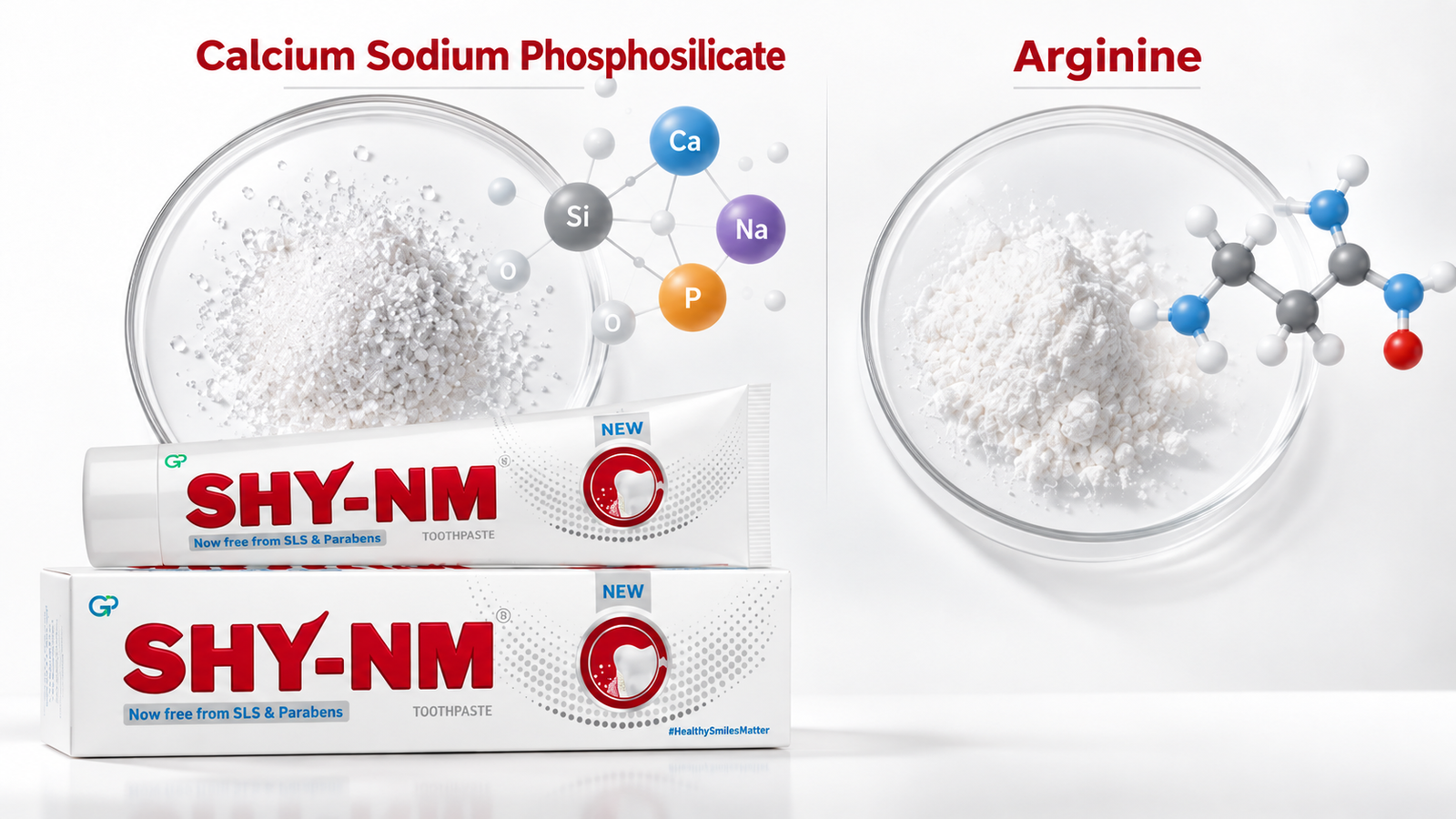
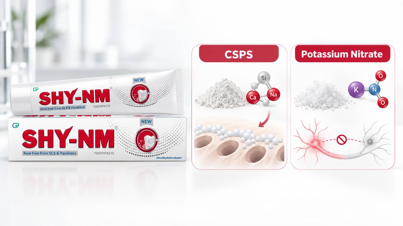
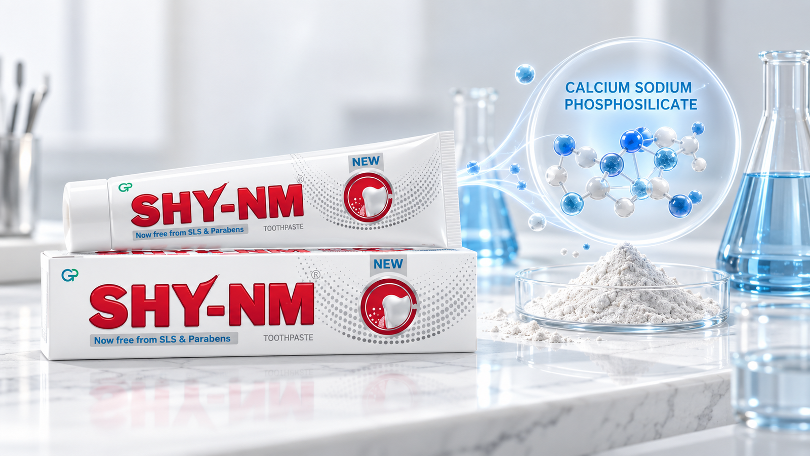
Calcium sodium phosphosilicate is a bioactive glass containing calcium, sodium, phosphorus and silicon.
When CSPS comes into contact with an aqueous environment, surface reactions can occur and ions can be released from the glass. These reactions create a mineral-rich environment around the material.
In dental applications, this behaviour has been investigated for its potential to support mineral deposition on tooth surfaces.
The 2017 Matsuyoshi study specifically examined whether a dentifrice containing 5% CSPS could promote remineralization of demineralized enamel.
Release of Calcium and Phosphate-Containing Species
The bioactive behaviour of CSPS is associated with ion release when the material interacts with an aqueous environment.
Calcium and phosphate-containing species are particularly relevant to mineral formation. Their availability can contribute to the development of a mineral-rich environment at the tooth surface.
This mechanism is one reason calcium sodium phosphosilicate has attracted interest as a bioactive material in dentistry.
However, it is important to distinguish the proposed material mechanism from a direct clinical claim. The presence of an ion-release mechanism does not by itself establish the extent of enamel remineralization that will occur in a patient’s mouth.
Development of a Mineral-Rich Surface Environment
As CSPS undergoes surface reactions, the local environment can become favourable to mineral deposition.
For enamel research, this is relevant because mineral recovery depends on the availability of ions and the conditions surrounding the enamel surface.
The interaction between CSPS, the surrounding fluid and the enamel surface is therefore an important part of understanding its potential remineralization behaviour.
Potential Contribution to Enamel Mineral Recovery
The proposed contribution of CSPS to enamel mineral recovery involves the formation and deposition of mineral phases at or within demineralized enamel.
The 2017 study investigated this process experimentally rather than assessing clinical caries outcomes. Its findings therefore provide evidence that CSPS-containing dentifrice can influence the remineralization process under controlled laboratory conditions, rather than establishing that the dentifrice clinically reverses enamel lesions in patients.
Evaluating Remineralization Using Optical Coherence Tomography
A key feature of the Matsuyoshi study was its use of optical coherence tomography, or OCT.
OCT is a non-invasive imaging technique that can produce cross-sectional images of semi-transparent biological structures. In dental research, it can be used to observe structural changes within enamel and monitor changes associated with demineralization and remineralization.
This makes OCT particularly useful for experimental studies where researchers need to monitor changes without destroying the specimen.
Why OCT Is Useful in Enamel Research
Traditional evaluation methods can require sectioning or otherwise altering a tooth specimen.
OCT provides an alternative approach for observing changes in enamel structure through optical signals. Changes in the intensity of back-scattered light can provide information about alterations within the enamel.
For remineralization research, this allows researchers to compare the optical characteristics of enamel before and after treatment.
The technique has been investigated as a method for monitoring enamel demineralization and remineralization, making it useful for laboratory studies of early enamel changes.
Study Methodology and Experimental Design
Matsuyoshi and colleagues used bovine incisors as the enamel specimens.
The specimens were prepared and divided into experimental groups. One group was subjected to an undersaturated lactic acid buffer at pH 4.75 for 10 minutes to create a demineralized enamel condition. The specimens were subsequently placed in artificial saliva at pH 7.0.
The study evaluated a dentifrice containing 5% calcium sodium phosphosilicate and used OCT to observe changes associated with enamel remineralization.
This experimental design allowed the researchers to examine the effect of the CSPS-containing dentifrice under controlled conditions.
It is important to note that the model was designed to investigate enamel behaviour in vitro. It did not reproduce all the biological factors present in the human oral cavity, such as natural saliva flow, dietary exposure, oral biofilm and individual patient behaviour.
Key Findings From the Study
The study reported that remineralization occurred when the enamel specimens were immersed in the dentifrice containing calcium sodium phosphosilicate.
The OCT observations provided evidence of changes associated with remineralization following exposure to the 5% CSPS dentifrice.
These findings support the potential of CSPS-containing dentifrice to contribute to mineral recovery in demineralized enamel under the experimental conditions used in the study.
At the same time, the findings should be interpreted within the scope of the experimental model. The study did not establish clinical effectiveness in humans, nor did it demonstrate that CSPS can independently reverse established dental caries.
Changes Observed After Treatment With 5% CSPS Dentifrice
The study focused specifically on a dentifrice containing 5% calcium sodium phosphosilicate.
Following treatment, the OCT observations indicated changes consistent with enamel remineralization. The authors concluded that the CSPS-containing dentifrice promoted remineralization of enamel under the conditions of the experiment.
This is particularly relevant to research into bioactive glass dentifrices because it provides experimental evidence that CSPS can interact with demineralized enamel in a way that is detectable through optical imaging.
The finding also adds to the broader scientific interest in CSPS as a bioactive material for dental applications.
Comparison With Demineralized Controls
The study included demineralized enamel specimens to establish a controlled experimental model.
The comparison allowed the researchers to observe changes in enamel following treatment with the CSPS-containing dentifrice rather than simply observing untreated enamel.
The resulting OCT observations supported the study’s conclusion that the 5% CSPS dentifrice promoted remineralization in the experimental model.
However, the comparison should not be interpreted as equivalent to a clinical comparison between CSPS toothpaste and another commercially available remineralizing treatment.
Clinical Interpretation for Dental Professionals
The Matsuyoshi study adds an important piece of experimental evidence to the discussion around calcium sodium phosphosilicate enamel remineralization.
It demonstrates that a 5% CSPS dentifrice can influence the remineralization of demineralized enamel under controlled laboratory conditions, with OCT used to monitor the changes.
For dental professionals, the significance lies in the biological plausibility and experimental observation of mineral recovery rather than in treating the study as proof of a clinical treatment effect.
Potential Role in Preventive Dentistry
The findings are relevant to preventive dentistry because early enamel mineral loss is an important stage at which non-invasive approaches may be considered.
A remineralizing dentifrice containing bioactive ingredients may be investigated as part of strategies designed to support enamel mineral recovery.
CSPS is of interest in this context because its bioactive glass chemistry allows it to interact with the surrounding fluid and release mineral ions.
However, preventive dentistry is multifactorial. Oral hygiene, fluoride exposure, dietary habits, saliva and individual caries risk all contribute to the balance between demineralization and remineralization.
Translating In-Vitro Findings Into Clinical Practice
One of the most important considerations when interpreting this research is the difference between laboratory findings and clinical outcomes.
The Matsuyoshi study used bovine enamel specimens under controlled experimental conditions. It did not involve human participants and therefore cannot establish how the same treatment would perform across different patients in everyday oral conditions.
In-vitro findings can provide valuable evidence about mechanisms and material behaviour, but clinical research is required to determine effectiveness in patients.
This distinction is particularly important when discussing terms such as enamel mineral recovery or demineralized enamel treatment. The study supports the potential for remineralization under experimental conditions, but it should not be interpreted as evidence that CSPS-containing toothpaste can repair all forms of enamel damage or reverse established cavities.
Study Limitations and Future Research
The study provides useful experimental evidence, but several limitations need to be considered.
First, the study was conducted using bovine enamel specimens, rather than human teeth in living participants. Although bovine enamel is widely used in dental research, it is an experimental substitute and does not reproduce every characteristic of human enamel.
Second, the study was performed under controlled laboratory conditions. The oral cavity is considerably more complex, with continuously changing saliva, oral biofilm, dietary exposure, mechanical brushing and individual variations in oral health.
Third, the study examined a specific dentifrice containing 5% CSPS. Its findings should therefore not automatically be applied to every calcium sodium phosphosilicate formulation or every concentration.
Finally, OCT provides valuable information about optical and structural changes, but an OCT observation alone does not establish a long-term clinical outcome.
Subsequent research has continued to investigate CSPS-containing dentifrices using different experimental models. For example, a 2024 in-vitro study of deeper incipient carious lesions found increased mineral density, hardness and elastic modulus in groups treated with CSPS-containing dentifrice compared with distilled water, while also reporting that remineralization at the lesion surface was similar among the tested dentifrices.
These findings illustrate why continued research using complementary laboratory techniques and well-designed clinical studies is valuable.
Conclusion
The evidence from the 2017 optical coherence tomography study provides experimental support for the potential of calcium sodium phosphosilicate enamel remineralization.
A dentifrice containing 5% calcium sodium phosphosilicate was observed to promote remineralization of demineralized enamel specimens under controlled laboratory conditions. OCT provided a non-invasive method for observing the structural changes associated with this process.
From a scientific perspective, the study supports the interest in calcium sodium phosphosilicate as a bioactive glass for dental applications and as an ingredient worth investigating in remineralizing dentifrices.
For clinical interpretation, however, the evidence should remain appropriately qualified. This was an in-vitro study using bovine enamel, so the findings demonstrate experimental remineralization rather than direct clinical effectiveness in patients.
Taken together, the study provides a useful foundation for understanding how CSPS-containing dentifrices may interact with demineralized enamel and highlights the value of further research connecting laboratory observations with clinical outcomes.
Reference
Matsuyoshi S, Murayama R, Akiba S, Yabuki C, Takamizawa T, Kurokawa H, Miyazaki M. Enamel remineralization effect of a dentifrice containing calcium sodium phosphosilicate: an optical coherence tomography observation. Acta Odontologica Scandinavica. 2017;75(3):191–197. DOI: 10.1080/00016357.2016.1277588.